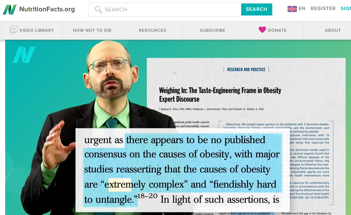
A friend for whom I have the greatest respect is a big fan of Michael Greger M.D. FACLM. To hear her talk, you’d think he walked on water. Personally, I didn’t much care for his style of presentation, and he seemed somewhat shallow on matters I know a bit about. But with several best-selling books and posts with catchy headlines such as “Does Cholesterol Size Matter?” and “Eat More Calories in the Morning than the Evening,” he has a legion of followers.
The announcement that Dr. Greger was going to do a series of video posts on obesity, really caught my attention. I’ve been interested in obesity for over 50 years. That’s how long I’ve known that MSG causes obesity. And I was excited that Dr. Greger might be going to share facts about the toxic effects of MSG. How MSG causes a-fib, migraine headache, fibromyalgia, skin rash, seizures, infertility, brain damage and more, not just that it causes obesity.
My excitement, however, was short-lived. Seems that even suggesting that MSG might cause obesity isn’t on Dr. Greger’s agenda. How do I know? Because I went to great lengths to contact him and suggest that MSG-related obesity was something he should look into. And on May 5, 2020 Christine Kestner, MS, CNS, LDN (Health Support Volunteer) responded:
“Hi, Adrienne Samuels! You can find everything on this site related to MSG here: https://nutritionfacts.org/topics/msg/ While it is true that this topic has not been updated in a while, a quick look at the lates research indicates that nothing has really changed in the last decade or so. We base our videos on the research, and not on industry influence. If you are aware of quality, peer-reviewed research that contradicts our positions, please share it with us.”
So, I did. I sent her pages of fully-referenced information. And then I waited. And waited. And then I sent a “You did get my letter, didn’t you?” note. And I’m still waiting.
Below is a copy of the material on MSG toxicity that Dr. Gregor ignored – or maybe Christine Kestner never showed it to him. Could be. Such is the power of the glutamate industry.
You’ll find the references for all this material at the end of the letter.
May 6, 2020
Thank you Christine,
The opportunity to provide accurate information about the toxicity of manufactured/processed free glutamate acid is much appreciated.
But first, two clarifications are in order. We generally speak of “MSG reactions,” but those reactions are actually caused by the Manufactured/processed free Glutamate (MfG) component of MSG. MfG is found in more than 40 food ingredients in addition to MSG. The animal studies listed below were done using MSG to inflict brain damage.
Second, glutamic acid will either be bound with other amino acids in protein or free. Bound glutamate does not cause brain damage or adverse reactions. Only glutamate in its free form causes brain damage and adverse reactions. This distinction is an important one, because failing to make it enables the fabrication of disinformation.
You said that a quick look at the latest research indicates that nothing has really changed in the last decade or so, but that is not entirely true.
I. MSG-induced brain damage. The seminal and definitive studies of MSG-inflicted brain damage were done in 1969 and the 1970s, and there is no need to replicate them.
In the late 60s, Olney became suspicious that obesity in mice, which was observed after neonatal mice were treated with L-glutamate for purposes of inducing and studying retinal pathology might be associated with hypothalamic lesions caused by L-glutamate treatment; and in 1969 he reported that L-glutamate treatment caused brain lesions, particularly acute neuronal necrosis in several regions of the developing brain of neonatal mice, and acute lesions in the brains of adult mice given 5 to 7 mg/g of glutamate subcutaneously (12). Research that followed confirmed that L-glutamate induces hypothalamic damage when given to immature animals after either subcutaneous (13-31) or oral (19,25-26,28,32-36) doses.
This work demonstrated that when there is a vulnerable target (a brain or portion of the brain that is unprotected or vulnerable to attack from toxins), and there is glutamic acid (glutamate) in quantity sufficient to cause it to become excitotoxic, glutamate fed in quantity to immature animals causes acute neuronal necrosis in several regions of the developing brain including the arcuate nucleus of the hypothalamus, followed by behavior disturbances and endocrine disruption which includes obesity and infertility.
A recent review suggests that glutamate/MSG passed to fetuses and neonates by pregnant and/or lactating women causes brain damage, disrupting the endocrine system (99).
It will be argued by agents of the glutamate industry that these studies of brain damage were animal studies not human studies, and that is true. But studies wherein possible toxins are fed to pregnant women and brains of their offspring are examined would certainly be questionable at best on ethical and moral grounds. Researchers rely heavily on animal studies to suggest solutions to problems of human dysfunction.
II. Industry’s unfounded claims of MSG safety
From 1968 until approximately 1980, Ajinomoto mounted a vigorous attack to refute the studies that demonstrated MSG-induced brain damage. Beginning in 1968 and throughout the 1970s, glutamate-industry agents mounted alleged replications of independently done glutamate-induced brain damage studies, but their procedures were different enough to guarantee that toxic doses had not been administered, and/or that all evidence that neurons had died would be obscured. Industry-sponsored researchers claimed to be replicating studies, but did not do so (5).
When it could no longer be denied that animal studies showed that MSG caused brain damage in infant animals – when researchers were using models of MSG-induced obesity to study abnormalities associated with excess glutamate — industry interests decreed that studies done on animals did not reflect the human condition and were, therefore, meaningless.
Industry-sponsored human studies followed in the 1980s. None were studies of brain damage.
III. Availability of sufficient potentially excitotoxic manufactured/processed free glutamate (MfG) in food and elsewhere to cause MfG to become excitotoxic (to kill brain cells)
Evidence of MSG-induced neonatal brain damage has not changed in the last four decades, but availability of sufficient glutamate in the U.S. food supply to cause that glutamate to become excitotoxic has.
Prior to 1957, the date that Ajinomoto reformulated MSG, the amount of free glutamate in the average diet had been unremarkable. But in 1957 production of the free glutamate that makes up the excitotoxic ingredient in MSG changed from extraction of glutamate from a protein source, a slow and costly method, to a method of bacterial fermentation which enabled virtually unlimited production of free glutamate and MSG (7), and the large amounts of glutamate needed to cause excitotoxicity became widely available.
Shortly thereafter, food manufacturers found that profits could be increased by producing other flavor-enhancing additives that contained free glutamate. Over the next two decades, the marketplace became flooded with manufactured/processed free glutamate in ingredients such as hydrolyzed proteins, yeast extracts, maltodextrin, soy protein isolate, and MSG (8). And ingredients that contained free glutamate became readily accessible.
There are no data on the amount of excitotoxic material in food. Analyses from Olney’s lab and others provided some insight into amounts of MSG in processed foods in the 1980s and 1990s (half a gram of MSG in certain canned soups, for example); and according to anecdotal reports from MSG-sensitive people, that would be enough to trigger an asthma attack or a migraine headache in some MSG-sensitive people. Reports from MSG-sensitive consumers also suggest that the amount of MfG in a single serving of processed food might be similar to that found in various cans of soup. None of this, however, speaks to the amount of MfG needed to produce either brain damage or adverse reactions.
Important to remember is the fact that it is not the amount of MfG in any one product that is pertinent to determining if there is sufficient MfG available to cause neonatal brain damage or adverse reactions. To cause neonatal brain damage, it is the amount of MfG consumed by a pregnant or lactating subject and passed to fetus and/or neonate that is relevant to determination of excitotoxicity.
IV. MSG-induced adverse reactions
There are few published reports of MSG-induced human adverse reactions. Funding for studies of the safety of MSG comes primarily from the glutamate industry, and only those industry-sponsored studies with negative results have been published.
Some years ago, Samuels compiled a list of studies wherein adverse reactions to MSG were noted (1-4, 175, 179-236). The article can be accessed at https://www.truthinlabeling.org/adverse.html .
No attempt has been made to identify all of the more recent studies. A PubMed search for “MSG-induced OR monosodium glutamate-induced AND toxicity” done on May 5, 2020 elicited 93 citations (https://www.ncbi.nlm.nih.gov/pubmed/?term=MSG-induced+OR+monosodium+glutamate-induced+AND+toxicity).
V. Warnings
By 1980, glutamate-associated disorders such as headaches, asthma, diabetes, muscle pain, atrial fibrillation, ischemia, trauma, seizures, stroke, Alzheimer’s disease, amyotrophic lateral sclerosis (ALS), Huntington’s disease, Parkinson’s disease, depression, multiple sclerosis, schizophrenia, obsessive-compulsive disorder (OCD), epilepsy, addiction, attention-deficit/hyperactivity disorder (ADHD), frontotemporal dementia, and autism were on the rise.
By and large, the glutamate in question was, and still is, glutamate from endogenous sources. The possible toxicity of glutamate from exogenous sources such as glutamate-containing flavor-enhancers has generally not being considered. But Olney and a few others have suggested that ingestion of free glutamate might play a role in producing the excess amounts of glutamate needed for endogenous glutamate to become excitotoxic (34-53).
VI. Suppression of information
The request to which I am responding was for quality peer-reviewed research that contradicts your positions. A list of those studies has been submitted with this letter.
Let me just mention that the videos you offered as the information on MSG safety came, directly or indirectly, from the glutamate-industry. The “update on MSG” is delivered by an unidentified person (as is “Is MSG Bad for You”) who speaks of scientific consensus and decades of research. The “scientific consensus” mentioned is the consensus of people brought together by Ajinomoto for the purpose of concluding that MSG is harmless. The “decades of research” were discussed earlier in this letter as negative studies that failed to demonstrate a clear and consistent relationship between MSG and adverse reactions. “Is MSG bad for you?” speaks only of consensus meetings. No sound scientific studies there. I would be happy to send you a link by email to my early notes on Williams and Woessner and on “the consensus meeting” should you have interest.
Equally important for you to appreciate are the studies that have been rigged by glutamate industry interests, and the tactics that have been used by glutamate-industry interests to promote sales of MSG. A 1999 published, peer-reviewed article speaks to that subject (101).
In addition, I have taken the liberty of enclosing the link to a file from my webpage titled “Designed for Deception.” Among other things, it details the tactics that Ajinomoto has used to rig its double-blind studies. (They stopped doing double-blind studies after we exposed the fact that they were lacing what they called “placebos” with aspartic acid, the excitotoxic amino acid used in aspartame. Aspartame and free aspartic acid cause the same brain damage and adverse reactions as those caused by MSG and free glutamic acid (32, 46, 102).
Additional reference
Neurobehav Toxicol. 1980 Summer;2(2):125-9.
Brain damage in mice from voluntary ingestion of glutamate and aspartate.
Olney JW, Labruyere J, de Gubareff T.
If there is anything else you would like me to provide to demonstrate that MSG kills brain cells and causes adverse reactions, please do not hesitate to contact me again.
Sincerely,
Adrienne Samuels, Ph.D.
Director
Truth in Labeling Campaign
Chicago, IL USA
truthlabeling@gmail.com
www.truthinlabeling.org
Reference used in this material
I. MSG-induced brain damage. The seminal and definitive studies of MSG-inflicted brain damage were done in 1969 and the 1970s, and there is no need to replicate them.
References
12. Olney JW. Brain lesions, obesity, and other disturbances in mice treated with monosodium glutamate. Science. 1969;164(880):719-721.
13. Olney JW, Ho OL, Rhee V. Cytotoxic effects of acidic and sulphur containing amino acids on the infant mouse central nervous system. Exp Brain Res. 1971;14(1):61-76.
14. Olney JW, Sharpe LG. Brain lesions in an infant rhesus monkey treated with monosodium glutamate. Science. 1969;166(903):386-388.
15. Snapir N, Robinzon B, Perek M. Brain damage in the male domestic fowl treated with monosodium glutamate. Poult Sci. 1971;50(5):1511-1514.
16. Perez VJ, Olney JW. Accumulation of glutamic acid in the arcuate nucleus of the hypothalamus of the infant mouse following subcutaneous administration of monosodium glutamate. J Neurochem. 1972;19(7):1777-1782.
17. Arees EA, Mayer J. Monosodium glutamate-induced brain lesions: electron microscopic examination. Science. 1970;170(957):549-550.
18. Everly JL. Light microscopy examination of monosodium glutamate induced lesions in the brain of fetal and neonatal rats. Anat Rec. 1971;169(2):312.
19. Olney JW. Glutamate-induced neuronal necrosis in the infant mouse hypothalamus. J Neuropathol Exp Neurol. 1971;30(1):75-90.
20. Lamperti A, Blaha G. The effects of neonatally-administered monosodium glutamate on the reproductive system of adult hamsters. Biol Reprod 1976;14(3):362-369.
21. Takasaki Y. Studies on brain lesion by administration of monosodium L-glutamate to mice. I. Brain lesions in infant mice caused by administration of monosodium L-glutamate. Toxicology. 1978;9(4):293-305
22. Holzwarth-McBride MA, Hurst EM, Knigge KM. Monosodium glutamate induced lesions of the arcuate nucleus. I. Endocrine deficiency and ultrastructure of the median eminence. Anat Rec. 1976;186(2):185-196.
23. Holzwarth-McBride MA, Sladek JR, Knigge KM. Monosodium glutamate induced lesions of the arcuate nucleus. II Fluorescence histochemistry of catecholamines. Anat Rec. 1976;186(2):197-205.
24. Paull WK, Lechan R. The median eminence of mice with a MSG induced arcuate lesion. Anat Rec. 1974;180(3):436.
25. Burde RM, Schainker B, Kayes J. Acute effect of oral and subcutaneous administration of monosodium glutamate on the arcuate nucleus of the hypothalamus in mice and rats. Nature. 1971;233(5314):58-60.
26. Olney JW, Sharpe LG, Feigin RD. Glutamate-induced brain damage in infant primates. J Neuropathol Exp Neurol. 1972;31(3):464-488.
27. Abraham R, Doughtery W, Goldberg L, Coulston F. The response of the hypothalamus to high doses of monosodium glutamate in mice and monkeys: cytochemistry and ultrastructural study of lysosomal changes. Exp Mol Pathol.1971;15(1):43-60.
28. Burde RM, Schainker B, Kayes J. Monosodium glutamate: necrosis of hypothalamic neurons in infant rats and mice following either oral or subcutaneous administration. J Neuropathol Exp Neurol. 1972;31(1):181.
29. Robinzon B, Snapir N, Perek M. Age dependent sensitivity to monosodium glutamate inducing brain damage in the chicken. Poult Sci. 1974;53(4):1539-1542.
30. Tafelski TJ. Effects of monosodium glutamate on the neuroendocrine axis of the hamster. Anat Rec. 1976;184(3):543-544.
31. Olney JW, Rhee V, DeGubareff T. Neurotoxic effects of glutamate on mouse area postrema. Brain Res. 1977;120(1):151-157.
32. Olney JW, Ho OL. Brain damage in infant mice following oral intake of glutamate, aspartate or cystine. Nature. 1970;227:609-611.
33. Lemkey-Johnston N, Reynolds WA. Nature and extent of brain lesions in mice related to ingestion of monosodium glutamate: a light and electron microscope study. J Neuropath Exp Neurol. 1974;33(1):74-97.
34. Takasaki, Y. Protective effect of mono- and disaccharides on glutamate-induced brain damage in mice. Toxicol Lett. 1979;4(3): 205-210.
35. Takasaki, Y. Protective effect of arginine, leucine, and preinjection of insulin on glutamate neurotoxicity in mice. Toxicol Lett. 1980;5(1):39-44.
36. Lemkey-Johnston, N, Reynolds WA. Nature and extent of brain lesions in mice related to ingestion of monosodium glutamate: a light and electron microscope study. J Neuropath Exp Neurol. 1974;33(1):74-97.
Reference
99. Samuels A. (2020). Dose dependent toxicity of glutamic acid: A review. International Journal of Food Properties. http://dx.doi.org/10.1080/10942912.2020.1733016
II. Industry’s unfounded claims of MSG safety
Reference
5. Samuels A. The toxicity/safety of processed free glutamic acid (MSG): a study in suppression of information. Accountability in Research.1999;6:259-310. https://www.truthinlabeling.org/assets/manuscript2.pdf Accessed 4/14/2020.
III. Availability of sufficient potentially excitotoxic manufactured/processed free glutamate (MfG) in food and elsewhere to cause MfG to become excitotoxic (to kill brain cells)
References
7. Hashimoto S. Discovery and History of Amino Acid Fermentation.
Adv Biochem Eng Biotechnol. 2017;159:15-34.
8. Sano C. History of glutamate production. Am J Clin Nutr. 2009;90(3):728S-732S
IV. MSG-induced adverse reactions
References
1. Reif-Lehrer, L. A questionnaire study of the prevalence of Chinese restaurant syndrome. Federation Proceedings 36:1617-1623,1977.
2. Kenney, RA and Tidball, CS Human susceptibility to oral monosodium L-glutamate. Am J Clin Nutr.25:140-146,1972.
3. Kerr, G.R., Wu-Lee, M., El-Lozy, M., McGandy, R., and Stare, F. Food-symptomatologyquestionnaires: risks of demand-bias questions and population-biased surveys. In: Glutamic Acid: Advances in Biochemistry and Physiology Filer, L. J., et al., Eds. New York: Raven Press, 1979.
4. Schaumburg, H.H., Byck, R, Gerstl, R, and Mashman, J.H. Monosodium L-glutamate: its pharmacology and role in the Chinese restaurant syndrome. Science 163:826-828,1969.
175. Kwok, R.H.M. The Chinese restaurant syndrome. Letter to the editor. N Engl J Med 278: 796, 1968.
179. Schaumburg, H. Chinese-restaurant Syndrome. N Engl J Med 278: 1122, 1968.
180. McCaghren, T.J. Chinese-restaurant syndrome. N Engl J Med 278: 1123, 1968.
181. Menken, M. Chinese-restaurant syndrome. N Engl J Med 278, 1123, 1968.
182. Migden, W. Chinese-restaurant syndrome. N Engl J Med 278: 1123, 1968.
183. Rath, J. Chinese-restaurant syndrome. N Engl J Med 278: 1123, 1968.
184. Beron, E.L. Chinese-restaurant syndrome. N Engl J Med 278: 1123, 1968.
185. Kandall, S.R. Chinese-restaurant syndrome. N Engl J Med 278: 1123, 1968.
186. Gordon, M.E., Chinese-restaurant syndrome. N Engl J Med 278: 1123-1124, 1968.
187. Rose, E.K. Chinese-restaurant syndrome. N Engl J Med 278: 1123, 1968.
188. Davies, N.E. Chinese-restaurant syndrome. N Engl J Med 278: 1124, 1968.
189. Schaumburg, H.H. and Byck, R. Sin cib-syn: accent on glutamate. N Engl J Med 279: 105, 1968.
190. Ambos, M., Leavitt, N.R., Marmorek, L., and Wolschina, S.B. Sin cib-syn: accent on glutamate. N Engl J Med 279: 105, 1968.
191. Schaumburg, H.H., Byck, R., Gerstl, R., and Mashman, J.H. Monosodium L-glutamate: its pharmacology and role in the Chinese restaurant syndrome. Science 163: 826-828, 1969.
192. Upton, A.R.M., and Barrows, H.S. Chinese-restaurant syndrome recurrence. N Engl J Med 286: 893-894, 1972
213. Gann, D. Ventricular tachycardia in a patient with the “Chinese restaurant syndrome.” Southern Medical J 70: 879-880, 1977.
214. Asnes, R.S. Chinese restaurant syndrome in an infant. Clin Pediat 19: 705-706, 1980.
215. Cochran, J.W., and Cochran A.H. Monosodium glutamania: the Chinese restaurant syndrome revisited. JAMA 252: 899, 1984.
216. Freed, D.L.J. and Carter, R. Neuropathy due to monosodium glutamate intolerance. Annals of Allergy 48: 96-97, 1982.
217. Ratner, D., Esmel, E., and Shoshani, E. Adverse effects of monosodium glutamate: a diagnostic problem. Israel J Med Sci 20: 252-253, 1984.
218. Squire, E.N. Jr. Angio-oedema and monosodium glutamate. Lancet 988, 1987.
219. Pohl, R., Balon, R., and Berchou, R. Reaction t chicken nuggets in a patient taking an MAOI. Am J Psychiatry 145: 651, 1988.
220. Reif-Lehrer, L. and Stemmermann, M.B. Correspondence: Monosodium glutamate intolerance in children. N Engl J Med 293: 1204-1205, 1975.
221. Andermann, F., Vanasse, M., and Wolfe, L.S. Correspondence: Shuddering attacks in children: essential tremor and monosodium glutamate. N Engl J Med 295: 174, 1975.
222. Reif-Lehrer, L. Letter: A search for children with possible MSG intolerance. Pediatrics 58: 771-772, 1976.
223. Reif-Lehrer, L. A questionnaire study of the prevalence of chinese restaurant syndrome. Fed Proc36:1617-1623, 1977.
224. Reif-Lehrer, L. Possible significance of adverse reactions to glutamate in humans. Federation Proceedings 35: 2205-2211, 1976. 225. Colman, A.D. Possible psychiatric reactions to monosodium glutamate. N Engl J Med 299: 902, 1978.
226. Neumann, H.H. Soup? It may be hazardous to your health. Am Heart J 92:, 266, 1976.
227. Gore, M.E., and Salmon, P.R. Chinese restaurant syndrome: fact or fiction. Lancet 1(8162): 251, 1980.
228. Sauber, W.J. What is Chinese restaurant syndrome? Lancet 1(8170): 721-722, 1980.
229. Allen, D.J., and Baker, G.J. Chinese-restaurant asthma. N Engl J Med 305: 1154-1155, 1981.
230. Allen, D.H., Delohery, J., & Baker, G.J. Monosodium L-glutamate-induced asthma. Journal of Allergy and Clinical Immunology 80: No 4, 530-537, 1987.
231. Moneret-Vautrin, D.A. Monosodium glutamate – induced asthma: Study of the potential risk in 30 asthmatics and review of the literature. Allergic et Immunologie 19: No 1, 29-35, 1987.
232. Smith, J.D., Terpening, C.M., Schmidt, S.O.F., and Gums, J.G. Relief of fibromyalgia symptonsfollowoing discontinuation of dietary excitotoxins. The Annals of Pharmacoltherapy. 35: (6) 702-706.
233. Scopp, A.L. MSG and hydrolyzed vegetable protein induced headache: review and case studies. Headache. 31:107-110, 1991.
234. Martinez, F. et al. Neuroexcitatory amino acid levels in plasma and cerebrospinal fluid during migraine attacks. Cephalalgia. 13: 89-93, 1993.
235. Scopp, A. Personal communication. June 17, 2002.
236. He K, Zhao L, Daviglus ML, et al. Association of Monosodium Glutamate Intake With Overweight in Chinese Adults: The INTERMAP Study. Obesity. 16(8): 1875-1880, 2008. Epub 2008 May 22.
V. Warnings
References
34. Hermanussen M, Tresguerres JA. Does high glutamate intake cause obesity? J Pediatr Endocrinol Metab. 2003;16(7):965-8.
35. Hermanussen M, García AP, Sunder M, Voigt M, Salazar V, Tresguerres JA. Obesity, voracity, and short stature: the impact of glutamate on the regulation of appetite. Eur J Clin Nutr. 2006;60(1):25-31.
36. Stover JF, Kempski OS. Glutamate-containing parenteral nutrition doubles plasma glutamate: a risk factor in neurosurgical patients with blood-brain barrier damage? Crit Care Med. 1999;27(10):2252-6.
37. Castrogiovanni D, Gaillard RC, Giovambattista A, Spinedi E. Neuroendocrine, metabolic, and immune functions during the acute phase response of inflammatory stress in monosodium Lglutamate-damaged, hyperadipose male rat. Neuroendocrinology. 2008;88(3):227-34.
38. Gill SS, Mueller RW, McGuire PF, Pulido OM. Potential target sites in peripheral tissues for excitatory neurotransmission and excitotoxicity. Toxicol Pathol. 2000;28(2):277-84.
39. Zautcke JL, Schwartz JA, Mueller EJ. Chinese restaurant syndrome: a review. Ann Emerg Med. 1986;15(10):1210-3.
40. Hermanussen M, Tresguerres JA. How much glutamate is toxic in paediatric parenteral nutrition? Acta Paediatr. 2005;94(1):16-9.
41. Nakanishi Y, Tsuneyama K, Fujimoto M, Salunga TL, Nomoto K, An JL, Takano Y, Iizuka S, Nagata M, Suzuki W, Shimada T, Aburada M, Nakano M, Selmi C, Gershwin ME. Monosodium glutamate (MSG): a villain and promoter of liver inflammation and dysplasia. J Autoimmun. 2008;30(1-2):42-50.
42. He K, Zhao L, Daviglus ML, Dyer AR, Van Horn L, Garside D, Zhu L, Guo D, Wu Y, Zhou B, Stamler J; INTERMAP Cooperative Research Group. Association of monosodium glutamate intake with overweight in Chinese adults: the INTERMAP Study. Obesity (Silver Spring). 2008;16(8):1875-80.
43. Niaz K, Zaplatic E, Spoor J. Extensive use of monosodium glutamate: A threat to public health? EXCLI J. 2018;17:273-278.
44. Olney JW. Excitotoxins in foods. Neurotoxicology. 1994;15(3):535-44.
45. Mondal M, Sarkar K, Nath PP, Paul G. Monosodium glutamate suppresses the female reproductive function by impairing the functions of vary and uterus in rat. Environ Toxicol. 2018;33(2):198-208.
46. Olney JW. Excitotoxic food additives–relevance of animal studies to human safety. Neurobehav Toxicol Teratol. 1984;6(6):455-62.
47. Onaolapo OJ, Onaolapo AY, Akanmu MA, Gbola O. Evidence of alterations in brain structure and antioxidant status following ‘low-dose’ monosodium glutamate ingestion. Pathophysiology. 2016;23(3):147-56.
48. Dixit SG, Rani P, Anand A, Khatri K, Chauhan R, Bharihoke V. To study the effect of monosodium glutamate on histomorphometry of cortex of kidney in adult albino rats. Ren Fail. 2014;36(2):266-70.
49. Zheng C, Yang D, Li Z, Xu Y. Toxicity of flavor enhancers to the oriental fruit fly, Bactrocera dorsalis (Hendel) (Diptera: Tephritidae). Ecotoxicology. 2018;27(5):619-626.
50. Olney JW. The toxic effects of glutamate and related compounds in the retina and the brain. Retina. 1982;2(4):341-59.
51. Olney JW. Excitatory neurotoxins as food additives: an evaluation of risk. Neurotoxicology. 1981;2(1):163-92.
52. Hashem HE, El-Din Safwat MD, Algaidi S. The effect of monosodium glutamate on the cerebellar cortex of male albino rats and the protective role of vitamin C (histological and immunohistochemical study). J Mol Histol. 2012;43(2):179-86.
53. Iamsaard S, Sukhorum W, Samrid R, Yimdee J, Kanla P, Chaisiwamongkol K, Hipkaeo W, Fongmoon D, Kondo H. The sensitivity of male rat reproductive organs to monosodium glutamate. Acta Med Acad. 2014;43(1):3-9.
VI. Suppression of information
Reference
101. Samuels A. The Toxicity/Safety of Processed Free Glutamic Acid (MSG): A study in Suppression of Information. Accountability in Research.1999(6):259-310 (https://bit.ly/2P4ICtd).


